| Jump to: Normal Exam Abnormal |
Sensory – Anatomy |
IntroductionClinically, there are 2 major somatosensory pathways that are examined. The first is the spinothalamic (ST) part of the anterolateral system and the second is the dorsal column-medial lemniscus (DCML) system. The principle sensory modalities for the ST system are pain and temperature. The principle sensory modalities for DCML system are vibratory, position sense and discriminatory or integrative sensation. |
|
|
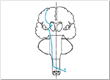
SpinothalamicThe anatomical pathways for the 2 major sensory systems is as follows: ST- the axons from the 1st order neuron located in the dorsal root ganglion enter the dorsal root entry zone and within several segments synapse with 2nd order neurons in the dorsal horn. Axons from the 2nd order neuron cross immediately via the ventral white commissure to the anterolateral quadrant of the spinal cord then ascend as the spinothalamic tract to the ventral posterior lateral nucleus (VPL) ofthe thalamus. The axons of the 3rd order neurons project to the postcentral gyrus or somatosensory cortex (there are also projections to the insular and anterior cingulate cortex but we are mainly focusing on the primary somatosensory cortex). |
|
|
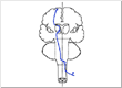
Dorsal Column-Medial LemniscusThe axons from the 1st order neurons located in the dorsal root ganglion enter the dorsal root entry zone and then ascend in the dorsal columns on the same side of the cord until they reach the 2nd order neurons in the medulla. Axons from the 2nd order neurons cross at the level of the medulla and then travel near the midline in the medial lemniscus. By the time the medial lemniscus reaches the rostral pons it has moved laterally and at this point it is in close proximity to the spinothalamic tract as it ascends to the VPL of the thalamus. The 3rd order neuron projects to the primary somatosensory cortex in the postcentral gyrus. |
|
|
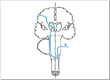
Trigeminal SystemThe trigeminal system is the somatosensory system for the face, which is clinically tested in the cranial nerve exam. For the trigeminal system it is important to remember that the descending tract of the trigeminal nerve, which serves pain and temperature, descends to the level of the upper cervical spinal cord and then axons from the 2nd order neurons cross over to the opposite side and ascend to the ventral posterior medial (VPM) nucleus of the thalamus. |
|
|

Level of CrossingThe following are important anatomical points to remember that have significant power in localizing lesions:
|
|
|

Location of Tracts
|
|
|

Trigeminal Crossing
|
|
|
Sensory DissociationThe above anatomical points translate into the following clinical findings:
|
|
|

Crossed Findings
|
|
|
Exam TestsThe ST is examined by testing:
The DCML is examined by testing:
|
|
|
Potential TrapsLight touch is represented in both the ST and DCML system so it is OK for sensory screening but not specific for either system. The sensory exam is perhaps the most subjective of the entire neurological exam so patient response can be difficult to interpret or at times be misleading. |
|
|
Clinical PearlsA sensory level is valuable in determining if there is spinal cord disease. Pain (sharp) is used to determine a sensory level. The sensory level on examination is usually 1-2 spinal cord segments below the actual spinal cord lesion. A sensory deficit from a spinal nerve lesion will be in a dermatome distribution. A sensory deficit from a peripheral nerve lesion will be in the distribution of that peripheral nerve. A sensory deficit from a polyneuropathy will have a stocking and glove distribution because the longest axons are the most affected. |
|
|
| Adapted, with permission from the University of Nebraska School of Medicine By Paul D. Larsen, M.D. and Suzanne S. Stensaas, Ph.D. |
|
|
| Jump to: Top of the Page Abnormal |
|
|
Sensory – Normal Exam |
Light TouchLight touch (thigmesthesia) is used as a screening test for touch. Both the spinothalamic and DCML systems serve this sensation so it is not specific for either one. A cotton tip applicator or fine hair brush is used. Select areas from different dermatomes and peripheral nerves and compare right versus left. |
|
|
Pain – Upper ExtremitiesPain is one of the principle sensory modalities of the spinothalamic system. The sharp end of a broken wooden cotton tip applicator can be used then discarded. It is important for the patient to be able to identify the sensation as sharp and then compare between dermatomes, distal versus proximal and right versus left for the upper extremities. |
|
|
Pain – Lower ExtremitiesPain is one of the principle sensory modalities of the spinothalamic system. The sharp end of a broken wooden cotton tip applicator can be used then discarded. It is important for the patient to be able to identify the sensation as sharp and then compare between dermatomes, distal versus proximal and right versus left for the lower extremities. |
|
|
TemperatureTemperature is the other sensory modality that is used to test the spinothalamic system. Tubes or vials of hot and cold water can be used but this is usually impractical. Using a tuning fork, which is normally perceived as cool or cold to the touch, compare between dermatomes and right versus left. |
|
|
VibratoryVibratory sensation (pallesthesia) is one of the sensory modalities of the DCML system. It is tested by using a 128 Hz tuning fork and placing the vibrating instrument over a bone or boney prominence. By varying the force of vibration and comparing the patient to yourself you can detect any deficits. Compare distal versus proximal and right versus left. |
|
|
Position SensePosition sense (proprioception), another DCML sensory modality, is tested by holding the most distal joint of a digit by its sides and moving it slightly up or down. First, demonstrate the test with the patient watching so they understand what is wanted then perform the test with their eyes closed. The patient should be able to detect 1 degree of movement of a finger and 2-3 degrees of movement of a toe. If the patient can’t accurately detect the distal movement then progressively test a more proximal joint until they can identify the movement correctly. |
|
|
Tactile MovementTactile movement as well as the remaining sensory tests are discriminatory sensory tests that examine cortical somatosensory (parietal lobe) function and require an intact DCML system. Tactile movement tests the patient’s ability to detect the direction of a 2-3 cm cutaneous stimulus. |
|
|
Two-Point DiscriminationTwo-point discrimination is tested by using calipers or a fashioned paper clip. The smallest and most dense sensory units are located in those areas that have the greatest somatosensory cortical representation. The patient should be able to recognize two-point separation of 2-4 mm on the lips and finger pads, 8-15 mm on the palms and 3-4 cm on the shins. |
|
|
GraphesthesiaGraphesthesia is the ability of the patient to identify characters that are written on the skin using a dull pointed object. The examiner demonstrates the test by writing single numbers on the palm of the hand while the patient is watching. The patient then closes their eyes and identifies numbers that are written by the examiner. |
|
|
StereognosisStereognosis is the ability to identify objects that are placed in the hand when the eyes are closed. The patient is given common objects and asked to identify them without looking at them. The inability to do this called astereognosis and indicates parietal lobe dysfunction. |
|
|
Double Simultaneous StimulationThe patient should be able to attend to and identify a tactile stimulus that is applied to both sides of the body at the same time. Double simultaneous stimulation (DSS) is tested by touching homologous parts of the body on one side, the other side or both sides at once with the patient identifying which side or if both sides are touched with their eyes closed. If the patient neglects one side on DSS (extinction or simultanagnosia) this indicates dysfunction of the contralateral posterior parietal lobe. |
|
|
Romberg TestThe Romberg test is a test of proprioception. This test is performed by asking the patient to stand, feet together with eyes open, then with eyes closed. The patient with significant proprioceptive loss will be able to stand still with eyes open because vision will compensate for the loss of position sense but will sway or fall with their eyes closed because they are unable to keep their balance. |
|
|
| Adapted, with permission from the University of Nebraska School of Medicine By Paul D. Larsen, M.D. and Suzanne S. Stensaas, Ph.D. |
|
|
| Jump to: Top of the Page Normal Exam |
|
|
Sensory – Abnormal |
Light TouchWith light touch the patient indicates that the perception of the stimulus is different over the left side of the face. The feeling has an abnormal quality to it described as different, uncomfortable or burning. This would be called paresthesia or dysesthesia. Light touch causing pain would be allodynia. |
|
|
Pain – Upper ExtremitiesA sharp wooden stick is used to delineate the area of decreased sharp sensation. There is loss over the ulnar side of the right hand as well as the ulnar aspect of the forearm but the arm is normal. This loss is constant with a C8-T1 dermatome distribution. |
|
|
Pain – Lower ExtremitiesThis patient has a sensory level at T3 with decreased pain sensation below the level including the leg. The sensory level is one to two spinal cord segment levels below the actual anatomical cord lesion because the spinothalamic axons ascend several spinal cord levels prior to crossing. The left sided T3 sensory level combined with this patient’s upper extremity sensory finding indicates a lesion of the right side of the spinal cord at the C8-T1 level. |
|
|
TemperatureThe patient is unable to distinguish the difference between a hot and cold test tube simultaneously applied to the ulnar side of the right hand and arm and the left leg. This deficit is in the same distribution as the pain deficit noted when testing sharp sensation. Pain and temperature sensation are tests for spinothalamic tract function. |
|
|
VibratoryVibratory sensation is decreased on the right great toe compared to the left. This could be due to a peripheral neuropathy but it also could be secondary to DCML deficit, which is actually the case for this patient. |
|
|
Position SenseThe patient makes more mistakes identifying the correct direction of toe movement on the right then left indicating a proprioceptive loss. For this patient this is secondary to a lesion effecting the dorsal column on the right side of the spinal cord. |
|
|
Tactile MovementWhen comparing left vs. right, the patient has more difficulty on the right side again indicating dorsal column dysfunction. If the dorsal column pathways are intact, then tactile movement is a sensitive test of parietal cortical function. |
|
|
Two-Point DiscriminationPatients with a lesion of the primary somatosensory cortex will have difficulty with two-point discrimination on the opposite side of the body. The peripheral nerve and DCML pathway must be intact for this test to be a test of parietal cortical function. |
|
|
GraphesthesiaThis patient has more difficulty identifying numbers written in the right hand than in the left hand. This is called agraphesthesia and is from a lesion of the somatosensory cortex in the left parietal lobe. |
|
|
StereognosisThe patient is asked to identify objects placed in both the right and left hand with his eyes closed. He knows that something is in his right hand but he is unable to identify it while he readily identifies the same object placed in the left hand. This is called astereognosis. The patient has a lesion involving the left parietal lobe. |
|
|
Double Simultaneous StimulationWhen the patient is touched on the right or left he correctly identifies the side touched but when both sides are touched simultaneously he neglects the stimulus on the right. This is extinction or simultanagnosia and indicates a lesion in his left parietal cortex. |
|
|
Romberg TestWith his eyes open, the patient is able to hold still but when his eyes are closed he sways and loses his balance. He has a significant loss of propioception. |
|
|
| Adapted, with permission from the University of Nebraska School of Medicine By Paul D. Larsen, M.D. and Suzanne S. Stensaas, Ph.D. |
|
|
| Jump to: Top of the Page Normal Exam Abnormal |
